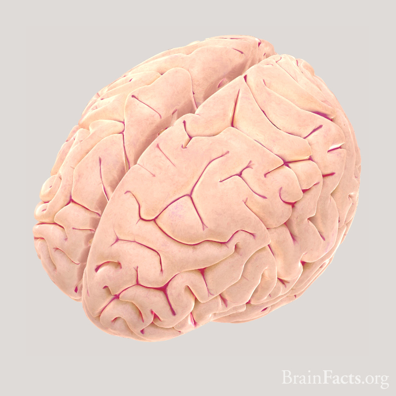
Superior view of human brain showing left and right cerebral hemispheres. Mapping by Jack Simpson.
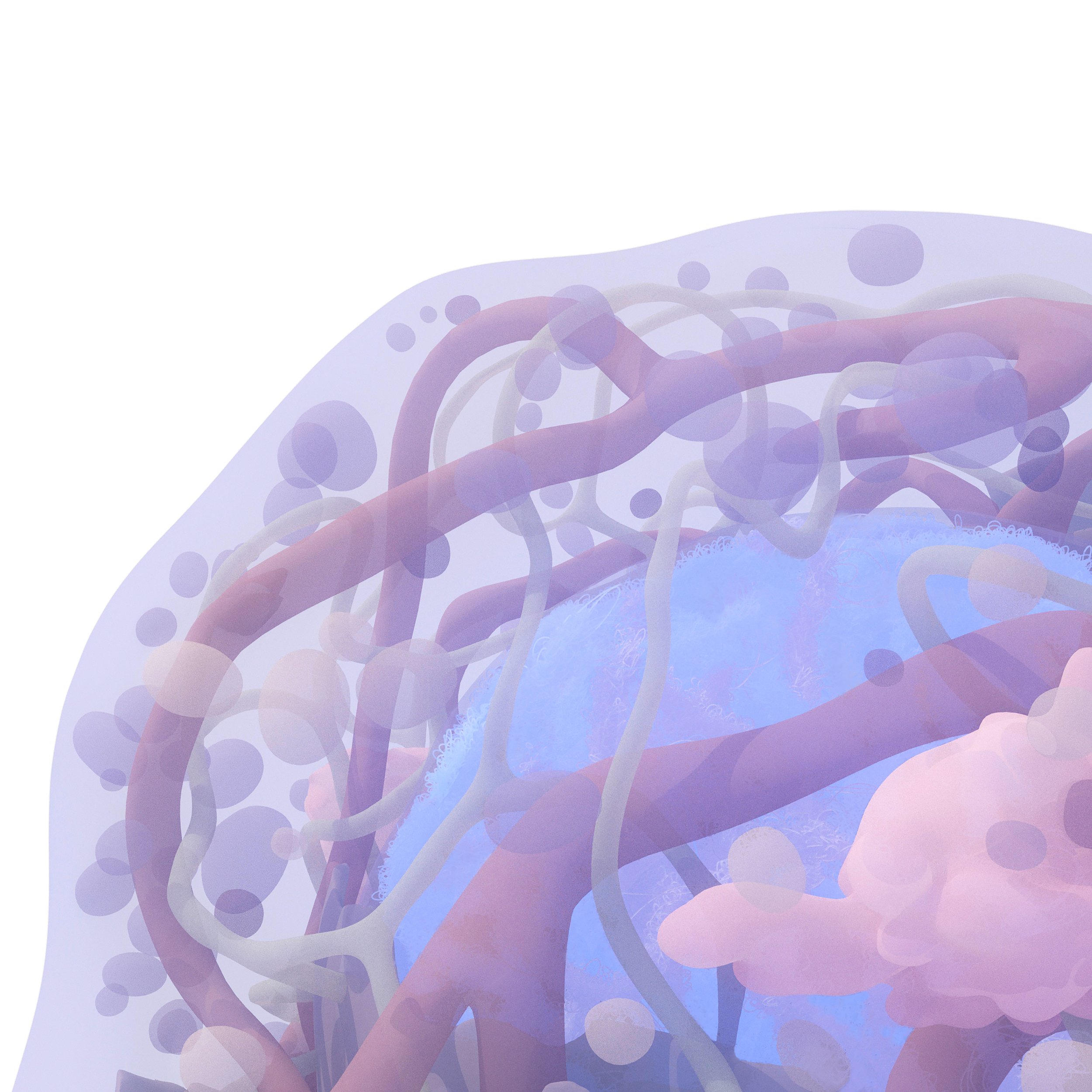
Stem cell featuring large nucleus, long tubular mitochondria, golgi apparatus, endosomes, lysosomes and microfilaments
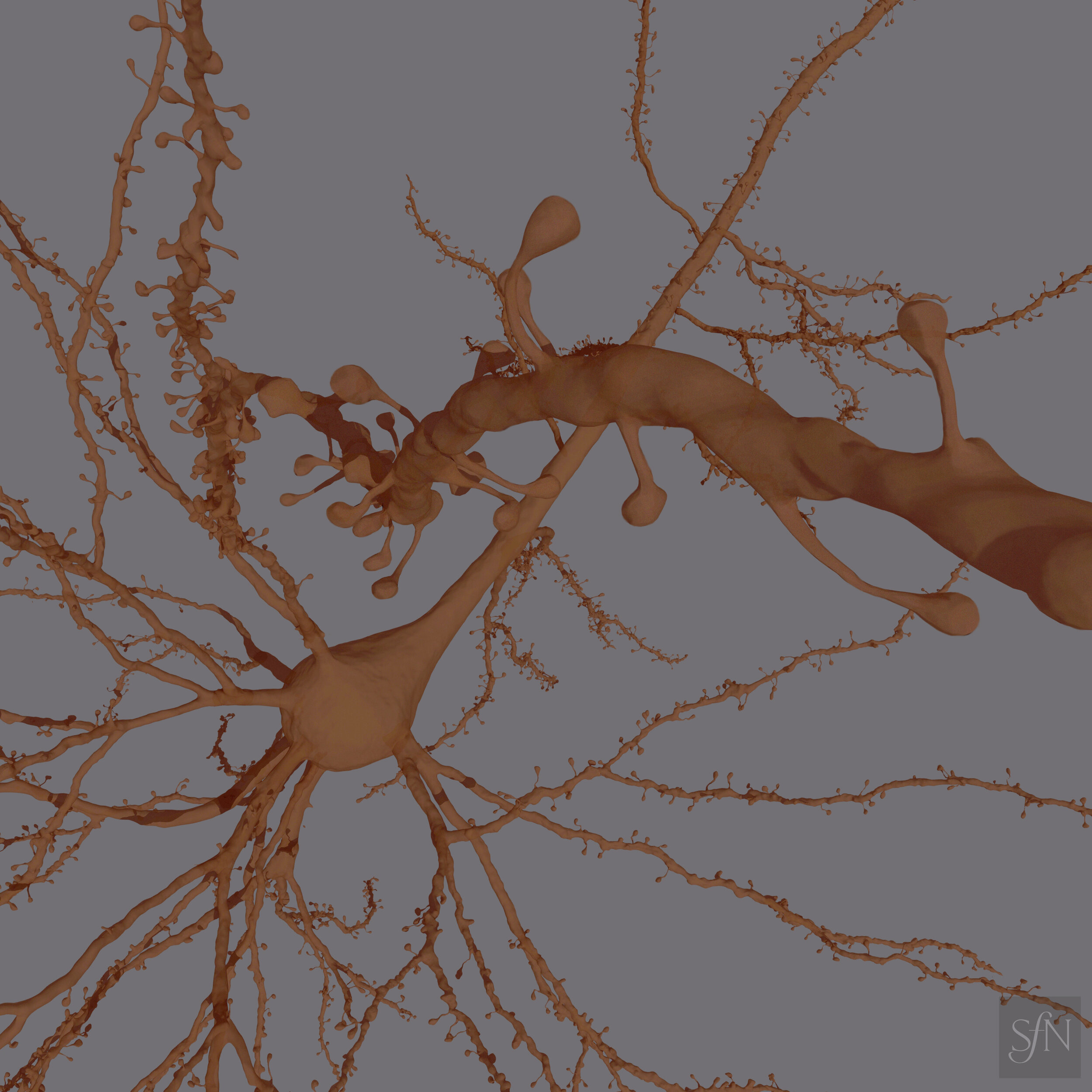
Neuron from the prefrontal cortex, layer 5. Model developed from scanned volume data provided by Peter Rogu and Dani Dumitriu, Columbia University.
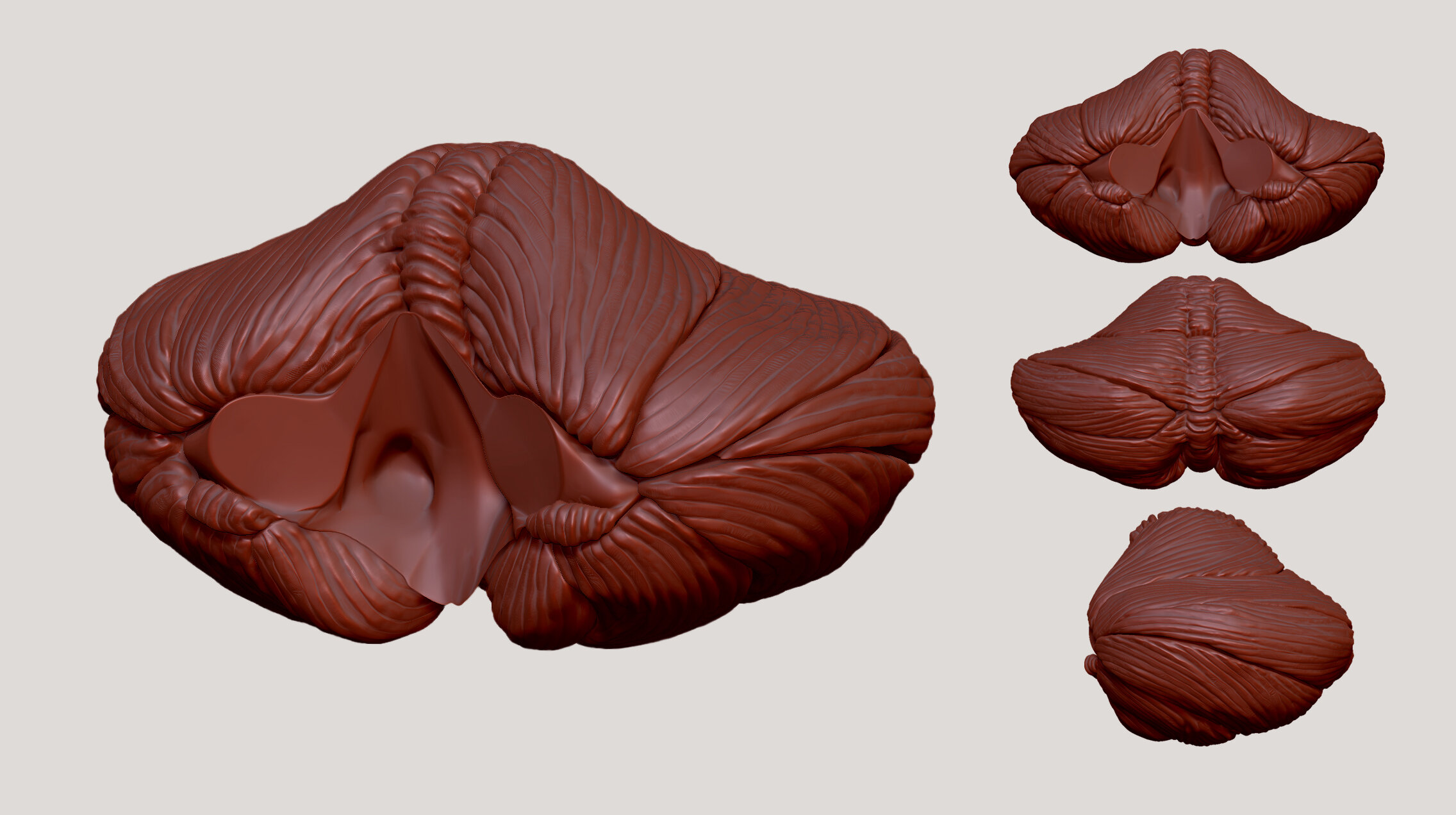
Multiple views of the cerebellum showing the different lobes divided by fissures.

Granular lymphocyte, proinflammatory white blood cell.
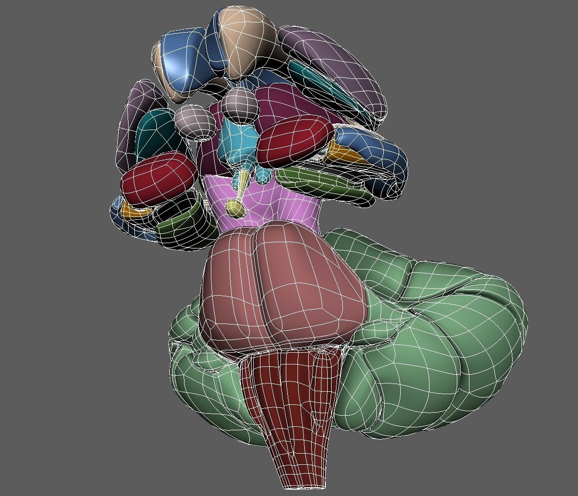
Subcortical brain structures including the limbic system, the brainstem and the cerebellum.
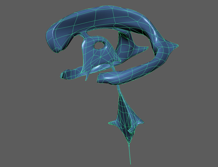
Cerebral spinal fluid filled ventricles found within the brain.
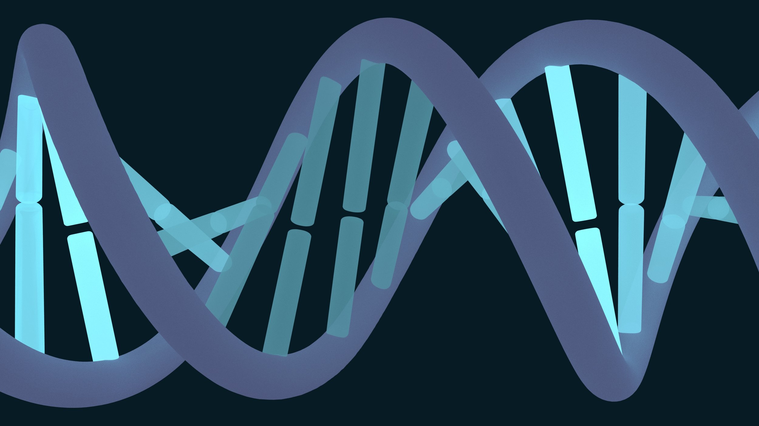
Double strand helical structure of DNA showing illuminated sequences of base pairs.

Immunoglobulin G is the most common antibody and has a long half life in circulation. Models derived from Protein Data Bank (PDB).
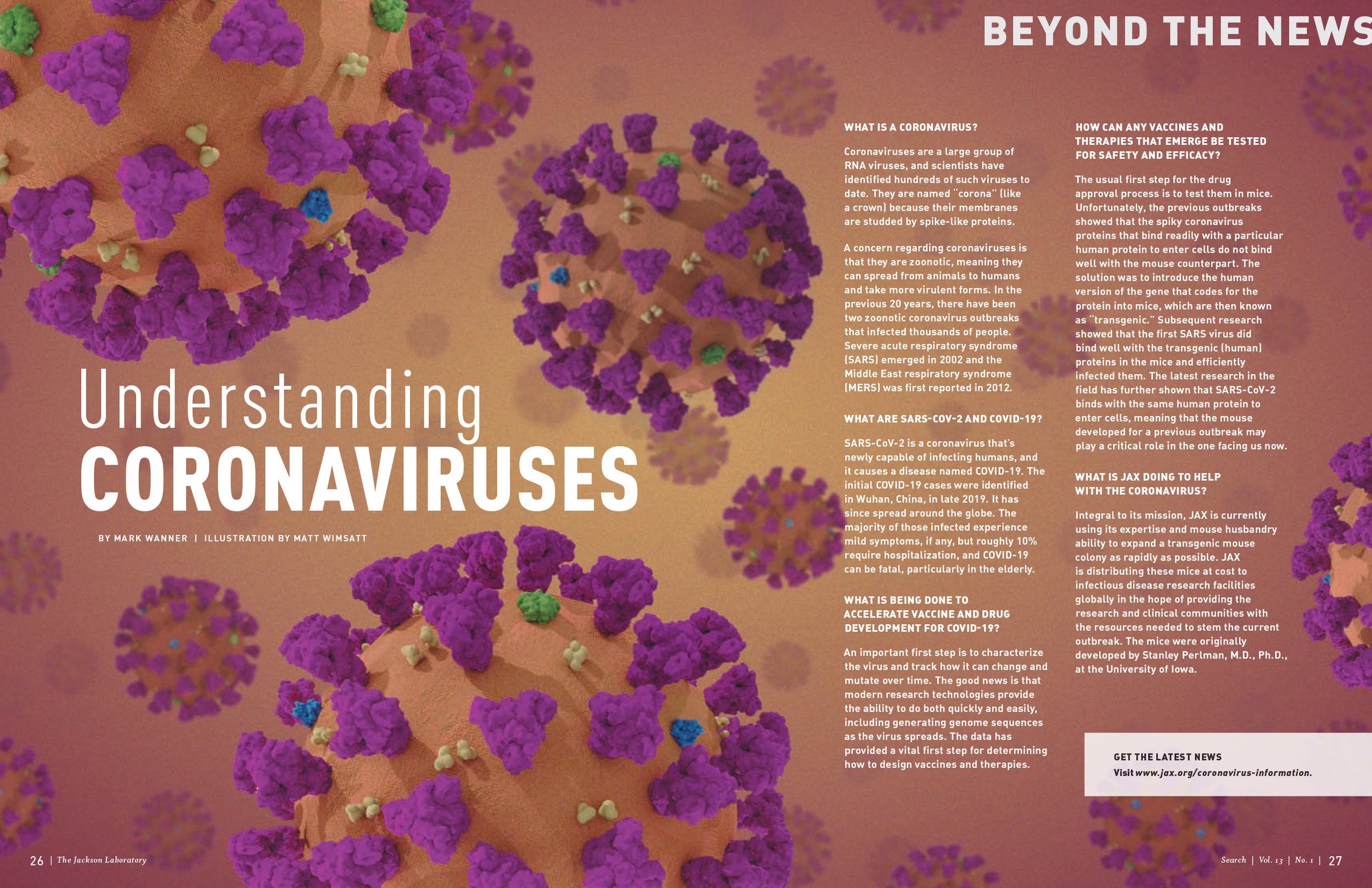
Magazine layout profiling the coronavirus structure featuring spike glycoproteins, hemagglutinin esterase, membrane proteins and envelope proteins. Molecular components sourced from the Protein Data Bank.
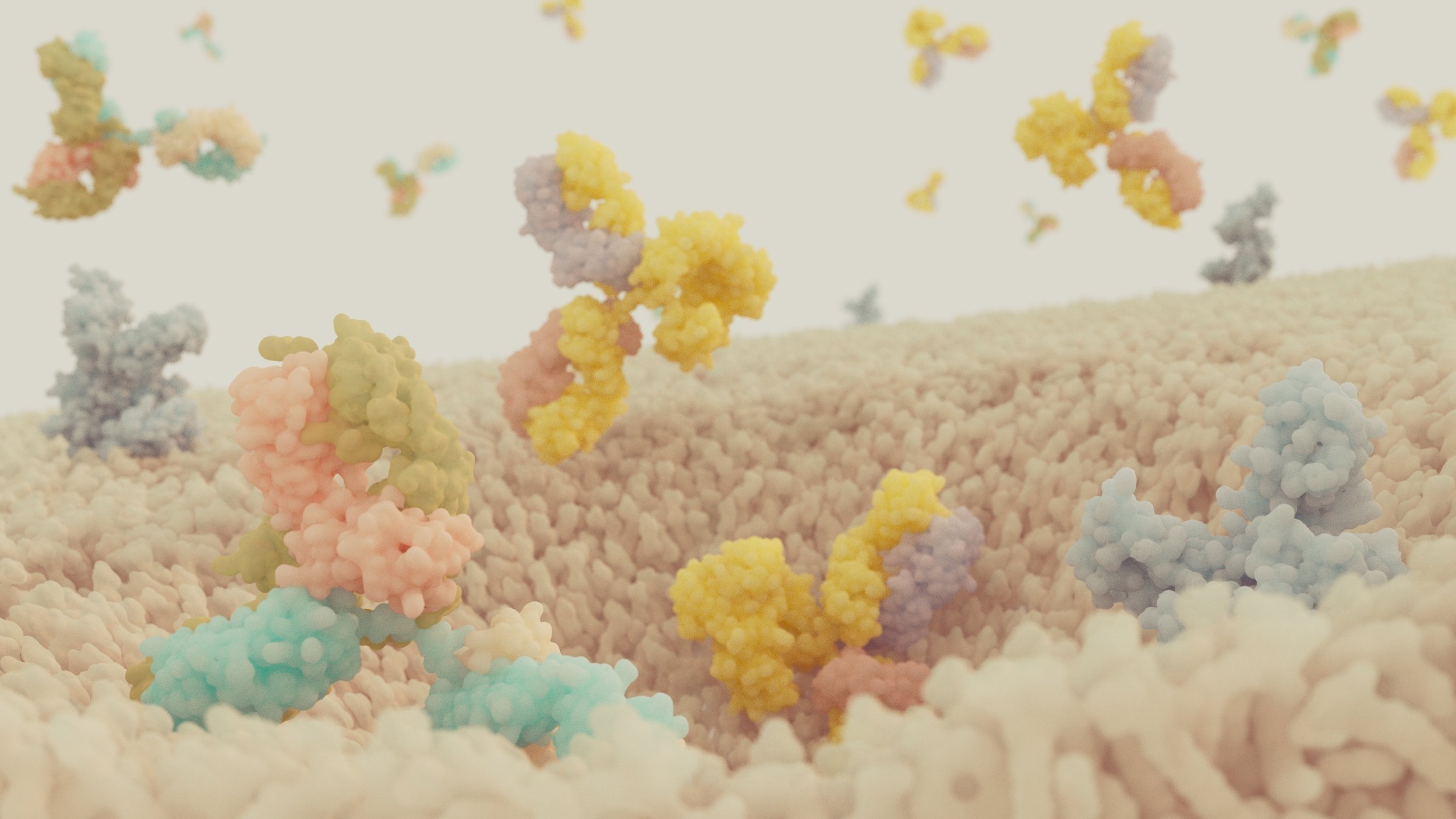
Scene on the surface of a cell showing FcRn receptors within the membrane with circulating IgG antibodies falling into a forming endosome. Models derived from Protein Data Bank (PDB).