Induced pluripotent stem cell (iPSC) featuring large nucleus and some organelles including long tubular mitochondria, golgi apparatus, endosomes, lysosomes and microfilaments. Model produced in Maya and ZBrush.

3D model of stem cell showing nucleus with surrounding organelles. Mitochondria branch out into a interconnected network around the nucleus. Rough endoplasmic reticulum wrap directly around the nucleus on one side with golgi flanking them. Free ribosomes circulate among lysosomes, peroxisomes and other small vesicles. Microtubules give the cell support as a long intertwining meshwork.


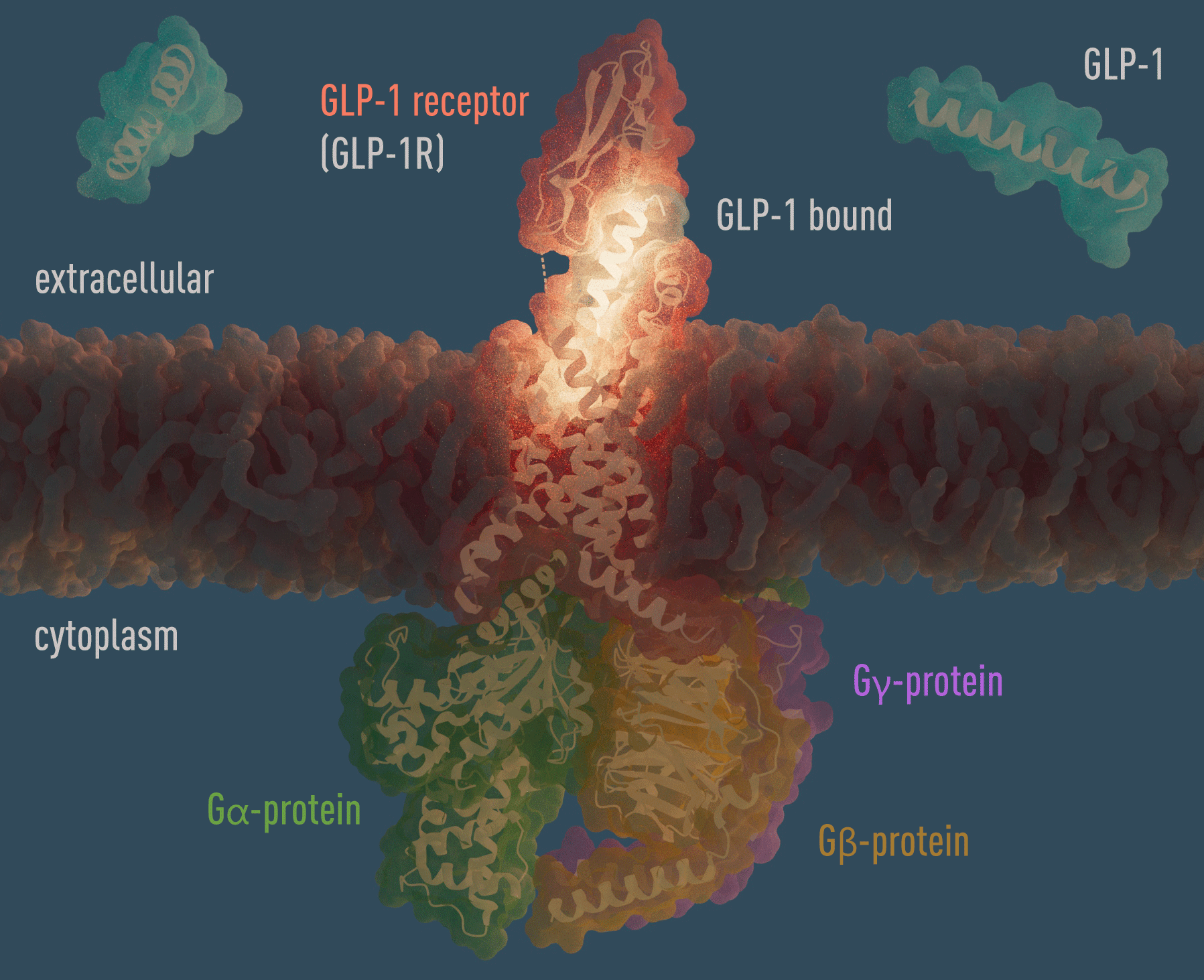
Apical cellular surface of the small intestine featuring Insulin, glucagon, GLP-1, ghrelin and leptin receptors, with glucose transporter GLUT5, and SGLT-1. Insulin (orange), leptin (pink), GLP-1 (turquoise), glucagon (blue), glucose (white), fructose (light orange) can be seen unbound in the extracellular space above. A GLP-1 agonist peptide can be seen binding a GLP-1 receptor. Sourced PDB entries: &STH, 2MFR, 1IRK, 8U4B, 2MFR, 2HIU, 6LML, 1GCN, 6X18, 3IOL, 7SL8, 4M56, 4YBQ, 4IW1, 7F9Y, 8DH8, 1AX8
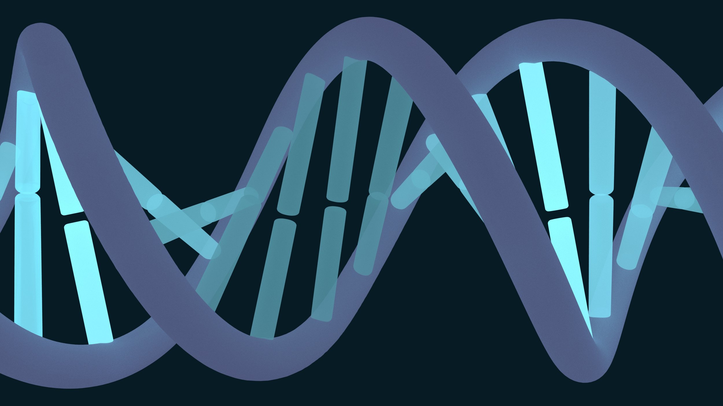
Double strand helical structure of DNA showing illuminated sequences of base pairs.

cross-sectional view within cellular endosome showing neonatal Fc receptor (FcRn) embedded within the endosomal bilipid membrane binding an IgG antibody and albumin. Other proteins and antibodies are also shown within the endosome. Only proteins bound to receptors will be recycled back out of the cell and not degraded.

Header images from the JAX Pulse monthly digital newsletter.
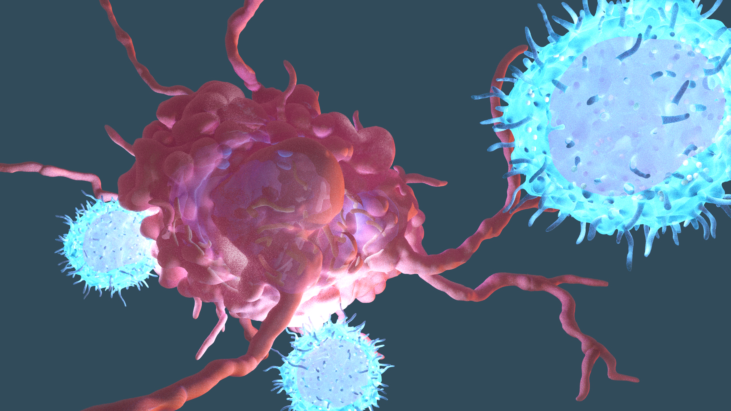
3D scene containing models of a cancer cell being confronted by activated cytotoxic T cells which are recognizing markers on the surface of the cancer cell. Cytotoxic T cells are part of the adaptive immune system.
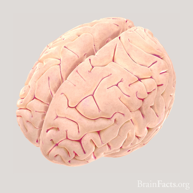
Superior view of human brain showing left and right cerebral hemispheres. Mapping by Jack Simpson.

Granular lymphocyte, proinflammatory white blood cell.
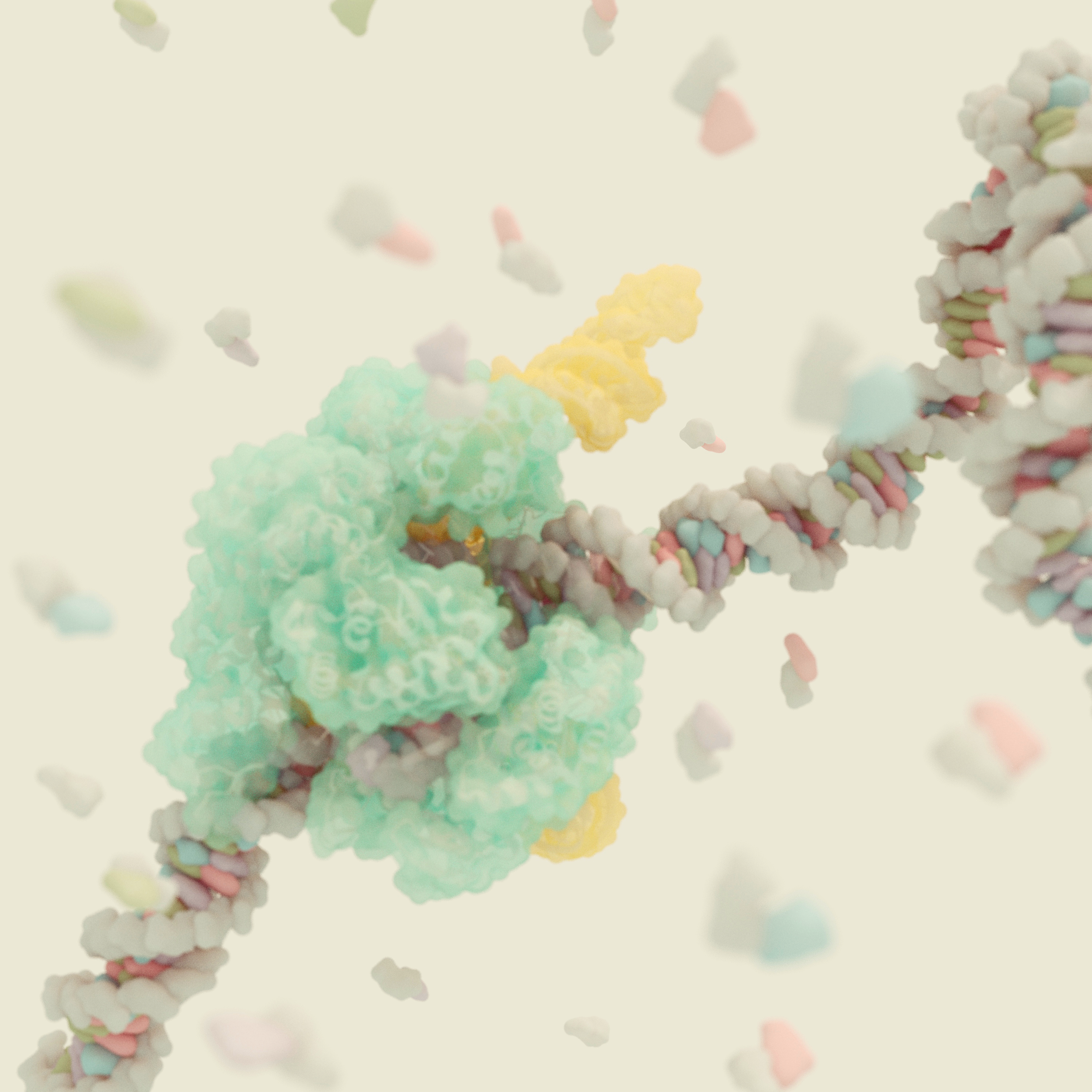
CRISPR Cas9 binding to dsDNA with nucleotides swirling about. Models resources: mMaya and PDB entry 5F9R

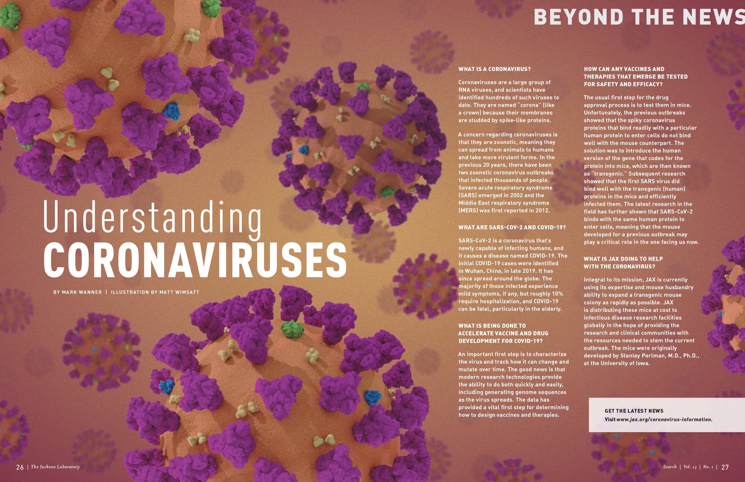
Magazine layout profiling the coronavirus structure featuring spike glycoproteins, hemagglutinin esterase, membrane proteins and envelope proteins. Molecular components sourced from the Protein Data Bank.
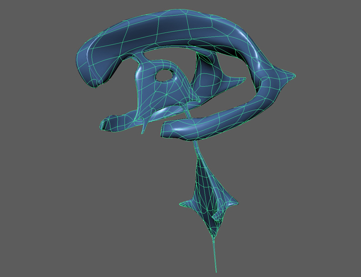
Cerebral spinal fluid filled ventricles found within the brain.
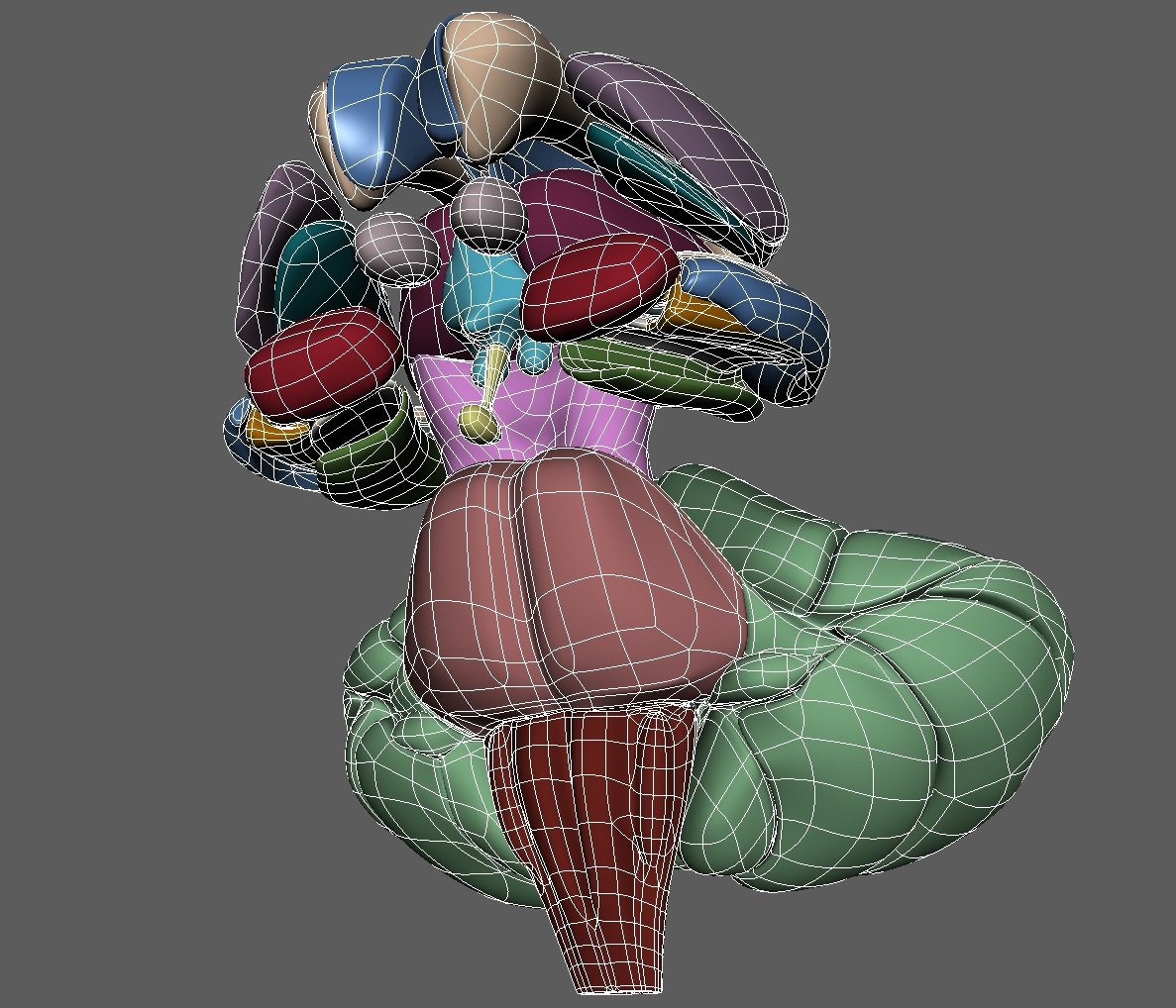
Subcortical brain structures including the limbic system, the brainstem and the cerebellum.

Conceptual behavioral assay simulation for mouse research.
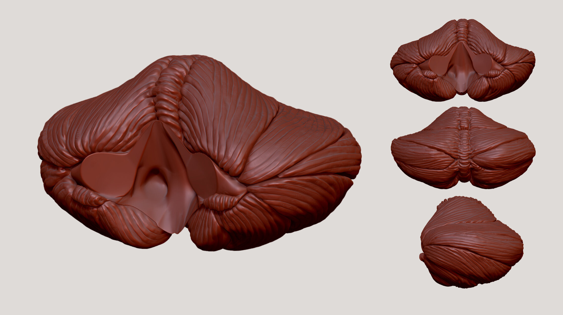
Multiple views of the cerebellum showing the different lobes divided by fissures.